Invertebrate
Zoology Microscopy
The compound light microscope focuses
visible light through a specimen. Different
regions of the object scatter the light differently, producing an image.
Three sets of lenses help generate the image.
The condenser lens focuses light through the
specimen. The objective lens receives light that has passed
through the specimen, generating an enlarged image.
The ocular lens, or eyepiece, magnifies the
image further.
All microscopes provide two types
of power—magnification and resolution (also called resolving
power). Magnification is the
ratio between the size of the image and the object. The total magnifying power is determined by
multiplying the power of the objective lens by the power of the ocular
lens. Resolution is a measure of the clarity of the image; it is the minimum distance two points can be separated and still be distinguished as two points. The function of the microscope is to enable
us to distinguish two points as separate even when they are so close together
that they appear as one to our unaided eye.
(Only when objects are farther than about 0.1 mm (100 mm) apart will your eye resolve them as two objects
rather than one, whereas a light microscope can resolve objects that are 0.1 to
0.2 mm apart.) The resolution or resolving power is
expressed as the relationship of two factors: the wavelength of light and the
numerical aperture.
Resolving power = |
___wavelength____ |
Because this is nothing more than
a fraction, and one that needs to be a very small number when divided out, the
wavelength must be small and the numerical aperture large. The reason we commonly use blue-violet
filters is because this end of the spectrum is the shortest wavelength that is
visible to the human eye.
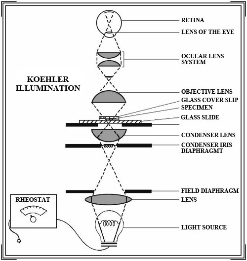
The light arriving at the specimen
is focused by the condenser lens from a round base to a point, thus having
the volume of a cone (see figure).
While it is unusual to think of light in volumetric terms; nevertheless,
that is the concept of numerical aperture.
It is a mathematical expression of the volume of light within the cone
of light delivered to the specimen and objective by the condenser.
The method used to ensure that this cone of light is at maximum volume
is called Kohler illumination.
a.
Begin the alignment at low illumination .
b. Use a 10X ocular (standard) and the 10X objective
c.
Place an easily visible test slide on the microscope.
d.
Open the condenser iris diaphragm and partially close
the field diaphragm.
e. Focus on the test slide. (With coarse focus control, lower objective
as far as it will go, while watching it to see that it does not crush the
slide. DO NOT look through the microscope
while lowering objective with coarse focusing knob. Looking through the microscope, raise
objective by means of coarse focus until object comes into view. Adjust the fine focus.)
f. Focus the image of the field diaphragm on
the test specimen by adjusting the height of the condenser (It will appear
polygonal when properly focused).
g. Center the image of the diaphragm by means of
the two setscrews located on the condenser.
h.
Open the field diaphragm until light fills the
field.
i.
When changing to 40X objective, partially close the
field diaphragm, and then repeat step f-h.
Microscopes should be par focal, so that only slight adjustments of fine
focus should be required when going from one objective to another.
a. Proper lighting is probably the most important requirement for good microscopy. Too much light is as bad as too little. Transparent objects are often clearer in reduced light (reduce light by closing down the iris diaphragm).
b. Focus with eyes relaxed. The image appears to your eye as though it is about 10 inches away, but your eye should be relaxed as though it is viewing an image in the distance. Look up periodically and train your eyes on something across the room. Then, as you keep your eyes relaxed, you should not have to readjust you focus very much when looking through the microscope. If you get a headache, chances are you are trying to look into the microscope rather than through it.
c. If you are using a binocular microscope, it
is important to (1) adjust the distance between the microscope’s oculars to
match the distance between your own eyes, and (2) adjust oculars for a sharp
focus.
d. Find the correct eye distance from the oculars, one that affords a full view of the field. Keep relaxed, hold your head steady and enjoy the view!
e.
If spots or smudges appear in the field of vision, it
may be dirt on the ocular, on the slide, or on the objective. To find out which, first rotate the ocular;
if the spots move, the ocular needs to be cleaned. Move the slide; if the spots move with it,
clean the slide. If after cleaning
ocular and slide the spots persist, it is probably a dirty objective lens. Use only lens paper to clean lenses. Never use alcohol, which will dissolve the
cement around the lens system. The slide
may be cleaned very gently with a damp Kim wipe. Some people may see “floaters” that drift
across the field of vision while viewing a brightly illuminated object. These are diffraction images of red blood
cell “ghosts” in the vitreous humor of the eye.
While annoying at first, you can usually learn to ignore them.
f.
Keep one hand on the fine adjustment and constantly
focus up and down. This allows you to
see detail through the depth of the object.
g. For oil immersion, first obtain a good image of the
desired area with the 40X objective.
Swing objective aside, and place a very small drop of immersion
oil over bright spot of light on the microscope slide. Swing 100X objective into place and adjust
fine focus. Always remove oil from the
100X objective with lens paper after use.
h. Be nice to your microscope. It must be kept dust free, so return it
carefully to its storage space when finished with it. Before putting it away, put the low-power
objective in place and raise the tube a little.
Be sure not to leave a slide on the stage.